|
|
|
|
| © W.P. Armstrong August 2007 |
|
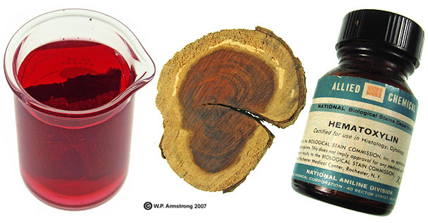
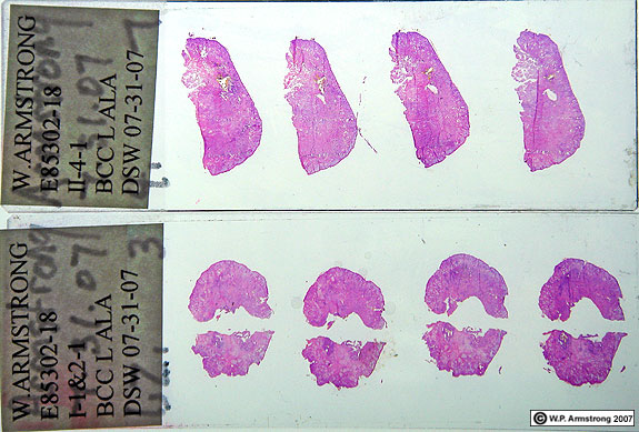
The following images are microscopic thin sections through the author's nose. The left ala was permeated by dense clusters of purple nuclei that show the tentacle-like, invasive growth of the tumorous basal cells. The tissue was stained with hematoxylin, a phenolic dye derived from the heartwood of logwood (Hematoxylum campechianum) native to Mexico & Central America. The microscopic images were taken at magnifications of 100x & 400x with a Sony V-3 digital camera. |

|
|


| Thin slice of left ala showing dense, dark-staining nuclei of tumor (morpheaform basal cell carcinoma). Several smaller sections of the tumor are also visible in this image. More images of the tumor are shown below. The color variations are due to slightly different exposures and different settings with Photoshop. |


|
A hair follicle and several sebaceous glands: .
|

| A hair follicle (middle right) and several sebaceous glands. Also called oil glands, the sebaceous glands secrete sebum, an oily substance that lubricates the hair and skin. If sebaceous glands fail to discharge, the secretions collect and form "whiteheads" or "blackheads." The darker color of blackheads is due to oxidized sebum. Image taken with Swift microscope & Sony V-3 Camera. Magnification 300x |

| A hair follicle (middle right) and several sebaceous glands. Also called oil glands, the sebaceous glands secrete sebum, an oily substance that lubricates the hair and skin. If sebaceous glands fail to discharge, the secretions collect and form "whiteheads" or "blackheads." The darker color of blackheads is due to oxidized sebum. Image taken with Olympus microscope & Sony V-3 Camera. Magnification 300x |